One of the most persistent and emotionally charged questions asked by individuals diagnosed with Type 1 Diabetes Mellitus (T1D), as well as their parents and family members, is simple yet profound: “Is Type 1 Diabetes genetic?”
When a child or young adult receives a T1D diagnosis, families often scour their medical genealogy looking for a history of the disease. In the vast majority of cases—over 85\%\text{ to }90\%—they find no prior family history at all. This creates an understandable paradox: How can a disease be genetic if nobody else in the family has it?
The short answer is that Type 1 Diabetes is a polygenic disease with strong hereditary susceptibility, but genetics alone rarely cause the condition. Instead, T1D develops through a complex biological dance: a genetically susceptible individual encounters environmental triggers that ignite an irreversible, cell-mediated autoimmune destruction of insulin-producing pancreatic beta cells.
This definitive guide, powered by MyDiaCalc, breaks down the human genome, the specific Human Leukocyte Antigen (HLA) complex, inheritance risks among relatives, and the vital difference between genetic predisposition and genetic destiny.
🚨 CRITICAL MEDICAL DISCLAIMER & LIMITATION OF LIABILITY (1/3): MyDiaCalc, its developers, authors, and operators DO NOT PROVIDE MEDICAL ADVICE. All information, risk percentages, genetic markers, and therapeutic insights presented in this article are intended strictly for educational, research, and informational purposes. MyDiaCalc EXPRESSLY DISCLAIMS ALL LIABILITY for any emotional distress, self-guided diagnostic interpretations, or clinical decisions resulting from the application of concepts discussed herein. Always consult with a board-certified endocrinologist or a licensed genetic counselor for personalized risk assessment and screening protocols.
1. Deconstructing the Genetics: Polygenic Susceptibility vs. Monogenic Diseases
To understand T1D inheritance, one must first distinguish between monogenic and polygenic genetic structures.
+———————————————————————–+
| MONOGENIC VS. POLYGENIC DIABETES PATTERNS |
+———————————————————————–+
| MONOGENIC DIABETES (e.g., MODY, Neonatal Diabetes) |
| └── Single gene mutation (e.g., GCK, HNF1A) |
| └── Direct Autosomal Dominant inheritance (50% pass-through risk) |
| └── Genetics = Direct Cause |
| |
| POLYGENIC DIABETES (Type 1 Diabetes Mellitus) |
| ├── Over 60+ distinct genomic loci implicated (HLA + Non-HLA) |
| ├── Requires additive effect of multiple risk alleles |
| └── Genetics = Susceptibility / Susceptibility Threshold |
+———————————————————————–+
Unlike disorders caused by a single mutated gene (such as Cystic Fibrosis or Maturity-Onset Diabetes of the Young – MODY), Type 1 Diabetes is polygenic. It requires a combination of risk alleles across dozens of genetic loci. Having these risk genes increases your “susceptibility bucket,” but environmental factors determine whether that bucket overflows into full-blown clinical autoimmunity.
Genome-Wide Association Studies (GWAS) Findings:
Modern genomic sequencing has identified more than 60 to 70 genetic loci associated with T1D risk. These genes do not code for “defective insulin”; rather, they govern how the immune system educates itself, recognizes foreign invaders, and identifies self-tissues.
2. The Primary Genetic Driver: The Human Leukocyte Antigen (HLA) Complex
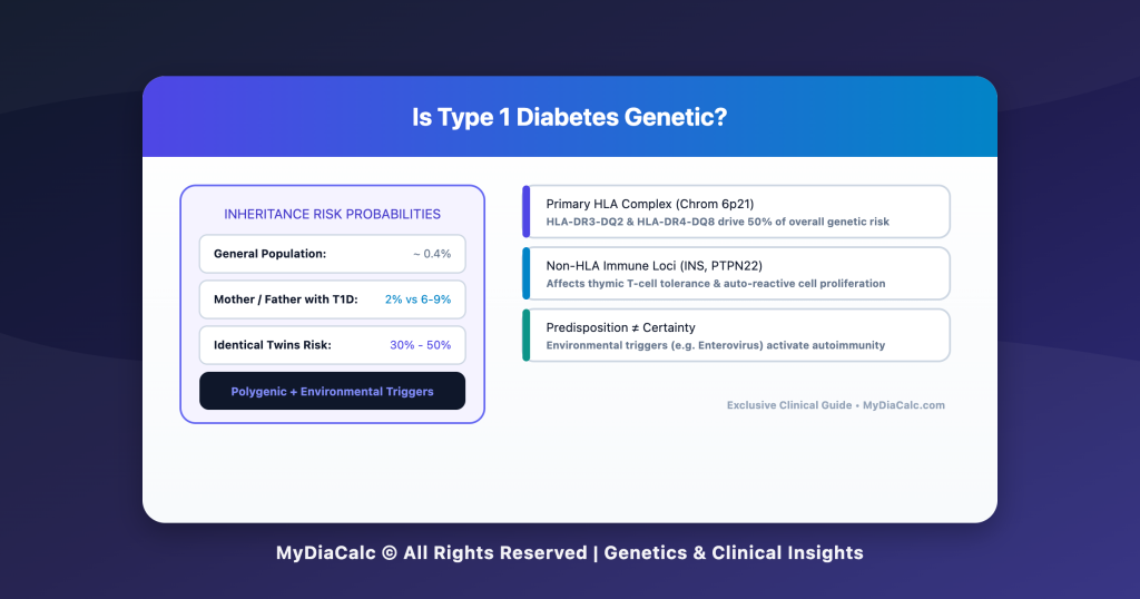
While dozens of genes play minor roles, 50% of the genetic risk for Type 1 Diabetes resides in a single cluster of genes located on Chromosome 6p21: the Human Leukocyte Antigen (HLA) region, specifically the Major Histocompatibility Complex (MHC) Class II genes.
+———————————–+
| Chromosome 6p21 (HLA Region) |
+———————————–+
|
+———————–+———————–+
| |
v v
+———————–+ +———————–+
| HLA-DR3 / DQ2 | | HLA-DR4 / DQ8 |
| (DRB1*0301-DQB1*0201) | | (DRB1*0401-DQB1*0302) |
+———————–+ +———————–+
| |
+———————–+———————–+
|
v
+———————————–+
| DR3/DR4 Heterozygote Combination |
| Highest Genetic Risk for T1D |
+———————————–+
How HLA Genes Function in Autoimmunity:
- Antigen Presentation: HLA Class II molecules (DR, DQ, DP) act as chemical “mangers” on the surface of Antigen-Presenting Cells (APCs). Their job is to hold peptide fragments and present them to T-helper lymphocytes.
- The High-Risk Conformational Pocket: In individuals with specific amino acid variants in the HLA-DQB1 chain (such as lacking an aspartic acid at position 57), the antigen-binding pocket binds fragments of pancreatic insulin and islet proteins in an unusual orientation.
- Loss of Immune Tolerance: T-cells incorrectly perceive these pancreatic self-proteins as dangerous foreign pathogens (like viruses), triggering an auto-reactive T-cell attack against the beta cells inside the Islets of Langerhans.
High-Risk Alleles at a Glance:
- HLA-DR3-DQ2 and HLA-DR4-DQ8 are present in roughly 90% of all children diagnosed with Type 1 Diabetes, compared to only 20% to 30% of the general population.
- The Synergistic Effect: Carrying both high-risk haplotypes (a DR3/DR4 heterozygote) creates the absolute highest genetic predisposition, elevating lifetime T1D risk significantly compared to the baseline population risk of 0.4\%.
3. Beyond HLA: Non-HLA Genetic Risk Factors
Although the HLA region dominates the risk profile, non-HLA genes account for the remaining half of genetic susceptibility. These genes primarily regulate T-cell signaling thresholds, central immune tolerance in the thymus, and innate inflammatory responses.
+———————————————————————–+
| KEY NON-HLA GENETIC CONTRIBUTORS TO T1D |
+———————————————————————–+
| Gene Locus | Primary Immunological Function |
+————+———————————————————-+
| INS | Regulates insulin expression in the thymus; educates |
| (11p15.5) | developing T-cells not to attack native insulin. |
+————+———————————————————-+
| PTPN22 | Encodes a phosphatase (LYP) that acts as a powerful |
| (1p13) | suppressor of T-cell receptor activation. |
+————+———————————————————-+
| CTLA4 | Encodes a critical checkpoint inhibitor receptor that |
| (2q33) | dampens auto-reactive T-cell proliferation. |
+————+———————————————————-+
| IL2RA | Encodes CD25 (IL-2 receptor alpha chain), essential for |
| (10p15) | the survival of Regulatory T-cells (Tregs). |
+———————————————————————–+
The Thymic Education Paradox (The INS Gene Variable):
The INS gene (insulin gene locus) variable number tandem repeat (VNTR) region dictates how much insulin is produced inside the thymus gland during early immune development:
- Protective Class III Alleles: Cause high insulin expression in the thymus. Developing T-cells that react to insulin are identified and destroyed before leaving the thymus (Central Tolerance).
- Predisposing Class I Alleles: Cause low insulin expression in the thymus. Auto-reactive T-cells escape destruction, enter circulation, and remain capable of attacking pancreatic beta cells later in life.
🚨 IMPORTANT MEDICAL DISCLAIMER & LIMITATION OF LIABILITY (2/3): Genetic Testing & Autoantibody Screening: Genetic risk markers (HLA typing) establish susceptibility, not active disease. MyDiaCalc ASSUMES ZERO LIABILITY for clinical misinterpretations, anxiety, or unprescribed medical interventions based on genetic risk evaluations. Active disease stage is measured exclusively via islet autoantibody panel testing (e.g., GAD65, IA-2, ZnT8, IAA) supervised by a physician.
4. Family Inheritance Risks: Quantifying the Probabilities
Because T1D involves polygenic traits alongside environmental triggers, the risk of inheriting the condition varies depending on which family member is affected.
+———————————————————————–+
| TYPE 1 DIABETES INHERITANCE RISK BY FAMILY MEMBER |
+———————————————————————–+
| Affected Family Member | Approximate Lifetime Risk |
+————————————–+——————————–+
| General Population (No family history)| ~ 0.4% (1 in 250) |
| Mother with T1D (Age at delivery > 28)| ~ 1% to 2% (1 in 100 to 50) |
| Mother with T1D (Age at delivery < 28)| ~ 4% (1 in 25) |
| Father with T1D | ~ 6% to 9% (1 in 16 to 11) |
| Both Parents with T1D | ~ 10% to 25% (up to 1 in 4) |
| Sibling with T1D | ~ 5% to 6% (1 in 20) |
| Non-Identical (Fraternal) Twin | ~ 5% to 7% (1 in 15) |
| Identical (Monozygotic) Twin | ~ 30% to 50% (1 in 2 to 3) |
+———————————————————————–+
Key Clinical Observations:
- The Paternal Predominance Paradox: Offspring of fathers with T1D have nearly double to triple the risk compared to offspring of mothers with T1D. The underlying mechanisms remain a subject of active research, involving genomic imprinting and maternal microchimerism during pregnancy.
- The Identical Twin Discordance: The fact that identical twins (who share 100\% of their DNA) have a concordance rate of only 30\%\text{ to }50\% is the single strongest proof that genetics alone do not dictate T1D development. If genetics were 100\% responsible, twin concordance would be 100\%.
The 15-15 Rule for Low Blood Sugar: A Clinical Step-by-Step Guide
5. The Environmental Trigger: What Activates the Genetic Gun?
Genetic susceptibility provides the “loaded gun,” but environmental triggers “pull the trigger.” In genetically at-risk infants, environmental factors are thought to initiate early islet autoimmunity (Stage 1 T1D).
+———————————–+
| Genetically Susceptible Host |
| (HLA-DR3/DR4, INS, PTPN22) |
+———————————–+
|
v
+———————————–+
| Environmental Triggers |
| (Enteroviruses, Gut Microbiome, |
| Early Diet, Vitamin D Def) |
+———————————–+
|
v
+———————————–+
| Stage 1: Autoimmunity |
| (≥2 Positive Islet Autoantibodies)|
+———————————–+
|
v
+———————————–+
| Stage 3: Clinical Diagnosis |
| (Dysglycemia + Beta-Cell Loss) |
+———————————–+
Leading Environmental Candidates:
- Enteroviral Infections (e.g., Coxsackievirus B): Enteroviruses display molecular mimicry with islet cell antigens (such as GAD65) and exhibit direct tropism for pancreatic beta cells, triggering inflammatory responses that breach self-tolerance.
- Gut Microbiome Alterations (Dysbiosis): Reduced gut bacterial diversity and compromised intestinal permeability (“leaky gut”) permit foreign food proteins and lipopolysaccharides to interact directly with gut-associated lymphoid tissue (GALT), activating auto-reactive T-cells.
- Early Infant Nutrition & Vitamin D: Early exposure to complex proteins or low vitamin D status during critical windows of immune maturation may alter regulatory T-cell development.
6. Clinical Screening & The Future of Prevention
Understanding the genetics of T1D has transformed medicine from reactive crisis management (treating DKA at onset) to proactive screening and early disease interception.
Disease Stages & Autoantibody Panels:
Genetics identify who is at risk, but autoantibodies track disease progression:
- Stage 1 T1D: Presence of \ge 2 islet autoantibodies (anti-GAD, IA-2A, ZnT8A, IAA) with normoglycemia.
- Stage 2 T1D: Autoantibodies present with asymptomatic dysglycemia.
- Stage 3 T1D: Clinical overt diabetes requiring exogenous insulin therapy.
Modern Immunotherapy Interceptions:
With the advent of disease-modifying therapies (such as FDA-approved monoclonal antibodies like Teplizumab), detecting genetic risk and autoantibodies early allows clinicians to delay the onset of Stage 3 clinical T1D by years, preserving endogenous beta-cell function and reducing acute onset complications.
Conclusion: Genetic Susceptibility, Not Genetic Destiny
Is Type 1 Diabetes genetic? Yes, but it is a disease of genetic predisposition rather than strict inheritance.
While inherited HLA risk alleles and non-HLA loci set the stage for immune dysregulation, environmental triggers and gut-immune dynamics dictate whether autoimmunity ultimately manifests. Understanding this polygenic architecture removes the misplaced guilt often felt by parents, opens the door for predictive autoantibody screening, and heralds a new era of immune-targeted preventive therapies.
Total Daily Dose & Basal Calculator
🚨 FINAL MEDICAL DISCLAIMER & LIMITATION OF LIABILITY (3/3): By utilizing MyDiaCalc, its calculators, tools, comparison charts, and educational guides, you explicitly acknowledge and agree that all therapeutic health decisions, glycemic interpretations, device selection choices, and clinical management decisions remain exclusively your personal responsibility or the responsibility of your licensed healthcare provider. MyDiaCalc, its authoring team, development staff, and affiliated entities SHALL NOT BE HELD LIABLE for any direct, indirect, incidental, consequential, or punitive damages resulting from the application or misuse of any calculations, guidelines, diagnostic benchmarks, or clinical concepts presented herein. Always work directly with your licensed endocrinologist or certified diabetes care specialist to establish safe individual treatment protocols.
Powered by MyDiaCalc © All Rights Reserved.