Lower extremity edema—commonly referred to as leg, ankle, or foot swelling—is a frequent and clinically significant presentation among individuals living with Type 1 and Type 2 Diabetes Mellitus. When patients notice their shoes feeling tighter or visible fluid accumulation above the ankles, a natural question arises: “Can diabetes cause leg swelling?”
The direct answer is yes. While diabetes itself does not directly “secrete” fluid into tissues, its chronic metabolic and microvascular complications systematically alter fluid dynamics, vascular permeability, renal clearance, and cardiac performance. Furthermore, several widely prescribed antidiabetic and antihypertensive medications can independently induce peripheral edema.
Identifying the root cause of leg swelling in diabetes is critical because peripheral edema ranges from benign fluid retention to early warning signs of end-stage organ dysfunction, such as Diabetic Nephropathy, Congestive Heart Failure (CHF), or Deep Vein Thrombosis (DVT).
This definitive clinical guide, powered by MyDiaCalc, breaks down the biological mechanisms behind diabetic fluid retention, systemic organ complications, drug-induced edema, diagnostic pathways, and evidence-based management strategies.
🚨 CRITICAL MEDICAL DISCLAIMER & LIMITATION OF LIABILITY (1/3): MyDiaCalc, its developers, authors, and operators DO NOT PROVIDE MEDICAL ADVICE. All clinical guidelines, diagnostic interpretations, and therapeutic concepts presented in this article are intended strictly for educational and informational purposes. MyDiaCalc EXPRESSLY DISCLAIMS ALL LIABILITY for any misdiagnoses, delayed medical interventions, acute heart failure exacerbations, or adverse drug events resulting from the application of concepts discussed herein. Always consult a board-certified endocrinologist, cardiologist, or nephrologist for formal evaluation of peripheral edema.
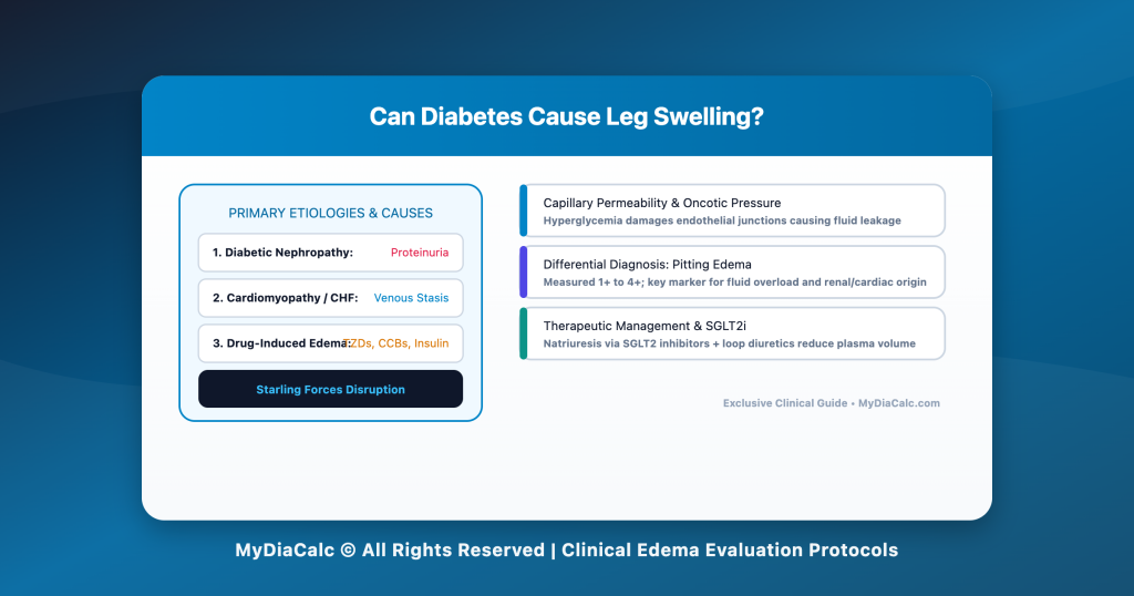
1. The Fluid Balance Equation: Starling Forces in Diabetes
To understand why diabetes leads to leg swelling, one must first examine the Starling Forces that govern fluid movement between capillaries and the interstitial tissue space.
+———————————————————————–+
| STARLING FORCES & EDEMA FORMATION |
+———————————————————————–+
| Capillary Hydrostatic Pressure (Pc) ───► Pushes fluid OUT to tissue |
| Interstitial Hydrostatic Pressure (Pi) ─► Pushes fluid INTO capillary |
| Capillary Oncotic Pressure (πc) ───────► Pulls fluid INTO capillary |
| Interstitial Oncotic Pressure (πi) ────► Pulls fluid OUT to tissue |
| |
| NET FLUID FLUX = Kf * [(Pc – Pi) – σ(πc – πi)] |
| * Edema occurs when Net Filtration EXCEEDS Lymphatic Drainage. |
+———————————————————————–+
Under normal physiological conditions, fluid filtered out of arterial capillaries is reabsorbed at the venous end or cleared by the lymphatic system. In diabetes, multiple biological disruptions skew this equilibrium:
- Increased Capillary Hydrostatic Pressure (P_c): Venous congestion from heart failure, venous insufficiency, or renal sodium retention elevates capillary pressure, forcing fluid out into tissues.
- Decreased Capillary Oncotic Pressure (\pi_c): Urinary protein loss (proteinuria) caused by diabetic kidney damage reduces plasma albumin, diminishing the “pulling force” that keeps fluid inside blood vessels.
- Increased Microvascular Permeability (K_f): Chronic hyperglycemia generates Advanced Glycation End-products (AGEs) and reactive oxygen species (ROS), which damage endothelial cell junctions and cause vessels to “leak” protein-rich fluid into the interstitium.
all-on-4 dental implants for diabetics
2. Major Causes of Leg Swelling in Diabetes
Leg swelling in diabetic patients is rarely caused by a single isolated factor. Rather, it usually stems from one of four major systemic categories:
+———————————————————————–+
| SYSTEMIC CAUSES OF DIABETIC LEG SWELLING |
+———————————————————————–+
| CATEGORY | PRIMARY BIOLOGICAL MECHANISM |
+—————————-+——————————————+
| 1. Diabetic Nephropathy | Glomerular damage -> Proteinuria -> |
| (Kidney Disease) | Hypoalbuminemia & Sodium Retention |
+—————————-+——————————————+
| 2. Cardiovascular Failure | Diabetic Cardiomyopathy / CAD -> |
| (Heart Failure) | Impaired ejection -> Venous Congestion |
+—————————-+——————————————+
| 3. Chronic Venous | Valve breakdown in leg veins -> |
| Insufficiency (CVI) | Venous pooling & Stasis Hyperpigmentation|
+—————————-+——————————————+
| 4. Medication-Induced | Thiazolidinediones (TZDs), CCBs, Insulin |
| Edema | renal sodium reabsorption & vasodilation |
+———————————————————————–+
A. Diabetic Nephropathy (Diabetic Kidney Disease – DKD)
Diabetic nephropathy is one of the most common causes of bilateral lower extremity edema. High blood glucose levels damage the delicate filtering units (glomeruli) within the kidneys over time.
- Microalbuminuria to Macroglobulinuria: As glomerular capillaries degrade, plasma proteins (primarily albumin) escape into the urine.
- Hypoalbuminemia: As serum albumin levels drop below normal thresholds (\le 3.5\text{ g/dL}), intravascular oncotic pressure plunges. Fluid leaks rapidly into dependent body regions, causing peripheral edema and, in severe cases, periorbital edema (swelling around the eyes).
- Renal Sodium Retention: Damaged kidneys fail to excrete sodium effectively, resulting in systemic volume overload.
B. Diabetic Cardiomyopathy & Heart Failure
People with diabetes face a 2 to 4 times higher risk of developing heart failure compared to non-diabetics.
- Ischemic Heart Disease: Accelerated atherosclerosis in coronary arteries impairs myocardial contraction.
- Diabetic Cardiomyopathy: Chronic hyperglycemia and insulin resistance cause direct myocardial fibrosis, stiffness, and diastolic dysfunction, even in the absence of coronary artery disease.
- Right-Sided vs. Left-Sided Failure: When the right side of the heart fails to pump blood effectively into the lungs, blood backs up into the systemic venous system, manifesting as severe, pitting leg edema and jugular venous distension (JVD).
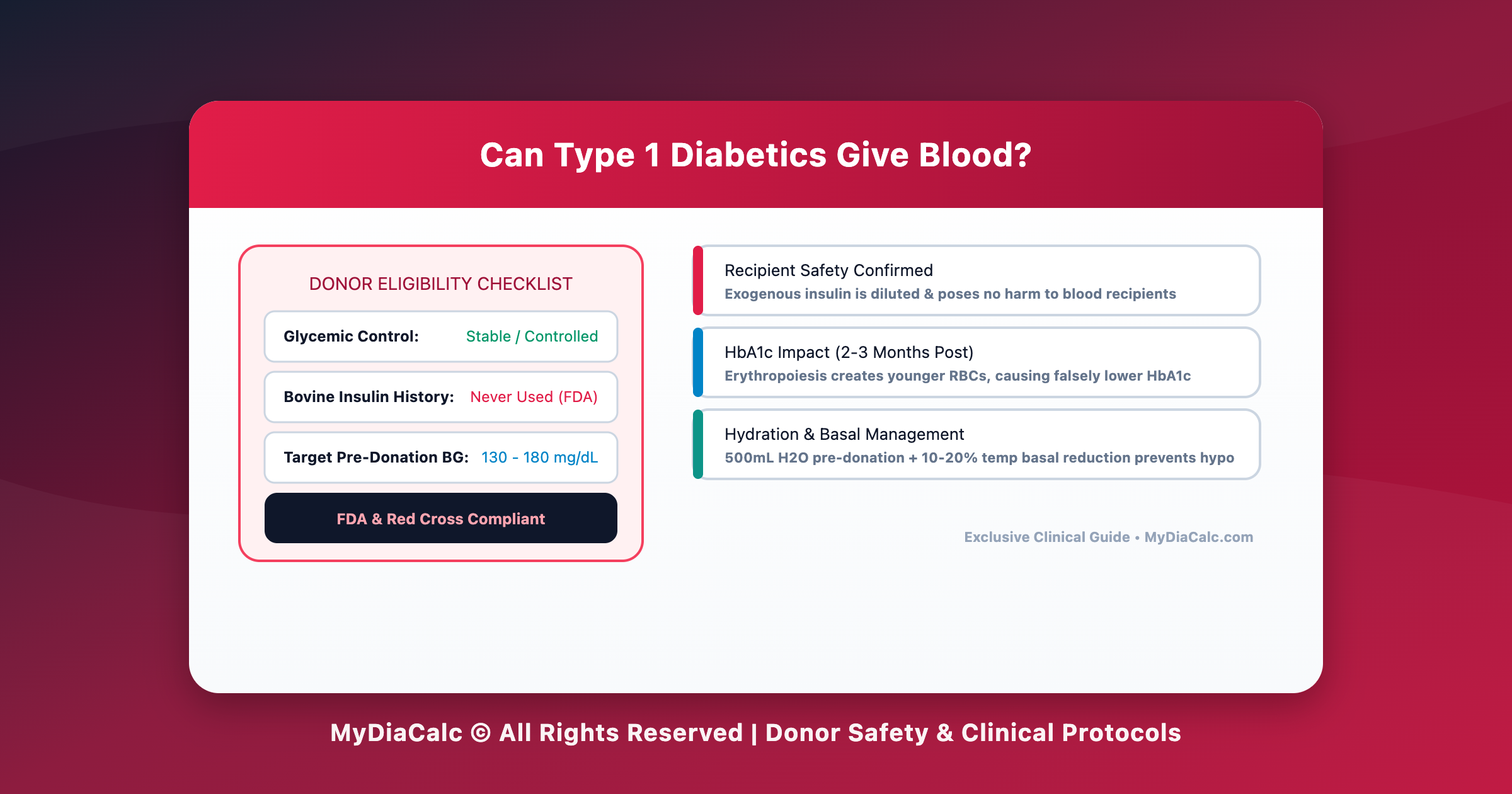
?can type 1 diabetics give blood
3. Vascular & Neurological Contributors: Venous Stasis & Neuropathy
Beyond renal and cardiac causes, localized vascular breakdown and nerve damage play prominent roles in chronic leg swelling.
+———————————–+
| Chronic Hyperglycemia |
+———————————–+
|
+———————–+———————–+
| |
v v
+———————–+ +———————–+
| Diabetic Neuropathy | | Endothelial Damage |
| (Loss of Muscle Pump | | (Venous Valve |
| & Vasomotor Tone) | | Incompetence) |
+———————–+ +———————–+
| |
+———————–+———————–+
|
v
+———————————–+
| Venous Pooling & Dependent Edema |
| (Ankles, Calves, Feet Bilaterally|
+———————————–+
Chronic Venous Insufficiency (CVI):
Diabetes impairs endothelial function and degrades the structural integrity of vein walls. The one-way valves inside deep and superficial leg veins become incompetent, preventing blood from flowing efficiently back up toward the heart. Blood pools in the lower legs under gravitational pull, increasing capillary pressure and causing chronic fluid extravasation.
Diabetic Autonomic & Peripheral Neuropathy:
- Impaired Vasomotor Tone: Damage to autonomic sympathetic nerves disrupts the normal constrictive response of blood vessels when standing, leading to vascular pooling.
- Loss of Calf Muscle Pump: Peripheral neuropathy causes muscle atrophy and gait abnormalities, diminishing the “calf muscle pump” action that normally compresses deep veins to propel blood upward during walking.
🚨 IMPORTANT MEDICAL DISCLAIMER & LIMITATION OF LIABILITY (2/3): Red-Flag Emergency Symptoms: Sudden, unilateral (one-sided) leg swelling accompanied by localized heat, redness, or calf pain may indicate a Deep Vein Thrombosis (DVT) or Cellulitis. MyDiaCalc ASSUMES ZERO LIABILITY for delayed emergency care. Seek immediate emergency medical attention if leg swelling is accompanied by shortness of breath, chest pain, or sudden asymmetric swelling.
4. Medication-Induced Edema in Diabetes
Managing diabetes often involves multi-drug regimens. Several classes of medications used to control blood glucose and blood pressure can cause fluid retention as a side effect.
+———————————————————————–+
| MEDICATIONS ASSOCIATED WITH LEG SWELLING |
+———————————————————————–+
| Drug Class | Common Examples | Mechanism of Edema |
+———————–+———————–+———————–+
| Thiazolidinediones | Pioglitazone, | Stimulates PPAR-γ in |
| (TZDs) | Rosiglitazone | renal collecting ducts|
| | | ──► Sodium reabsorption|
+———————–+———————–+———————–+
| Calcium Channel | Amlodipine, | Pre-capillary arteriolar|
| Blockers (CCBs) | Nifedipine | vasodilation ──► High |
| | | capillary pressure |
+———————–+———————–+———————–+
| Exogenous Insulin | Insulin Glargine, | Renal tubular sodium |
| Therapy | Lispro, Degludec | retention (Insulin |
| | | Edema Phenomenon) |
+———————–+———————–+———————–+
| Pregabalin / | Lyrica, | Central/peripheral |
| Gabapentin | Neurontin | vascular tone changes |
+———————————————————————–+
The TZD & Insulin Impact:
- TZDs (Pioglitazone): These PPAR-\gamma agonists directly enhance sodium reabsorption in the renal collecting tubules, increasing plasma volume. They are contraindicated in patients with NYHA Class III or IV heart failure.
- Insulin Edema: When initiating intensive insulin therapy in patients with chronic severe hyperglycemia, a sudden drop in blood glucose causes the kidneys to retain sodium and water rapidly—a transient condition known as insulin edema.
5. Differential Diagnosis: Pitting vs. Non-Pitting Edema
When evaluating leg swelling, clinicians distinguish between pitting and non-pitting edema to narrow down the underlying etiology.
+———————————–+
| Physical Examination of Edema |
| (Press thumb for 5 seconds) |
+———————————–+
|
+———————–+———————–+
| |
v v
+———————–+ +———————–+
| PITTING EDEMA | | NON-PITTING EDEMA |
| (Persistent Indentation| | (No Persistent Indent)|
+———————–+ +———————–+
| • Heart Failure | | • Lymphedema |
| • Renal Disease (DKD) | | • Advanced Thyroid |
| • Venous Stasis | | Disorders (Myxedema)|
| • Medication-Induced | | • Lipedema |
+———————–+ +———————–+
Measuring Pitting Severity:
- 1+ (2 mm depth): Mild indentation; disappears almost instantly.
- 2+ (4 mm depth): Moderate indentation; rebounds in 10 to 15 seconds.
- 3+ (6 mm depth): Deep pit; lasts more than 1 minute; noticeable swelling of lower leg.
- 4+ (8 mm depth): Very deep pit; persists for 2 to 5 minutes; gross distorted extremity.
6. Clinical Diagnostic & Management Framework
A structured clinical evaluation ensures early detection of high-risk conditions like renal failure or decompensated heart failure.
+———————————————————————–+
| DIAGNOSTIC & MANAGEMENT ACTION PLAN |
+———————————————————————–+
| CLINICAL WORKUP | • Urine Albumin-to-Creatinine Ratio (uACR). |
| | • Serum Creatinine & eGFR (Kidney Function). |
| | • B-type Natriuretic Peptide (BNP / NT-proBNP). |
| | • Echocardiogram (Assess Left Ventricular EF). |
| | • Venous Duplex Ultrasound (Rule out DVT/CVI). |
+———————+————————————————-+
| THERAPEUTIC STEP 1 | Glycemic Control: Optimize HbA1c to prevent |
| (Glycemic & Renal) | microvascular damage using SGLT2 inhibitors. |
+———————+————————————————-+
| THERAPEUTIC STEP 2 | Pharmacotherapy: Initiate Loop Diuretics |
| (Fluid Excretion) | (e.g., Furosemide) for volume overload. |
+———————+————————————————-+
| THERAPEUTIC STEP 3 | Non-Pharmacological: Compression stockings |
| (Mechanical) | (20-30 mmHg) + leg elevation above heart level. |
+———————————————————————–+
The Role of SGLT2 Inhibitors:
Modern cardiovascular-renal agents like Sodium-Glucose Cotransporter-2 (SGLT2) inhibitors (e.g., Empagliflozin, Dapagliflozin) offer dual therapeutic benefits: they promote urinary glucose and sodium excretion (natriuresis), reducing plasma volume, lowering capillary hydrostatic pressure, and protecting kidney/heart function simultaneously.
Conclusion: Early Detection Protects Organ Function
Can diabetes cause leg swelling? Yes, peripheral edema is a clear biological signal that diabetes is impacting vascular permeability, kidney clearance, cardiac performance, or medication tolerance.
Recognizing leg swelling early allows patients and healthcare providers to intervene before irreversible microvascular or macrovascular damage occurs. Through comprehensive clinical diagnostic panels, targeted pharmacotherapy (such as SGLT2 inhibitors and diuretics), and lifestyle modifications, fluid retention can be managed effectively—preserving long-term mobility and quality of life.
🚨 FINAL MEDICAL DISCLAIMER & LIMITATION OF LIABILITY (3/3): By utilizing MyDiaCalc, its calculators, tools, comparison charts, and educational guides, you explicitly acknowledge and agree that all therapeutic health decisions, glycemic interpretations, device selection choices, and clinical management decisions remain exclusively your personal responsibility or the responsibility of your licensed healthcare provider. MyDiaCalc, its authoring team, development staff, and affiliated entities SHALL NOT BE HELD LIABLE for any direct, indirect, incidental, consequential, or punitive damages resulting from the application or misuse of any calculations, guidelines, diagnostic benchmarks, or clinical concepts presented herein. Always work directly with your licensed endocrinologist, nephrologist, or cardiologist to establish safe individual treatment protocols.
Powered by MyDiaCalc © All Rights Reserved.